About CHRPE
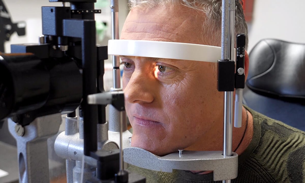
A flat, pigmented spot within the outer layer of the retina at the back of the eye is called a congenital hypertrophy of the retinal pigment epithelium (CHRPE). The pigmentation of the lesion can range from a light gray to black.CHRPE is a great example of why you should get your eyes checked at least once a year. You can't see the hypertrophic lesion or "spot" just by looking in your eyes. It can be detected in an eye exam by your primary optometrist, ophthalmologist, or retina specialist. This is a congenital condition meaning you are born with it, but it may go undetected until later in adulthood. In the vast majority of cases, CHRPE is a benign finding that never causes a problem with vision or life. Visit any of our locations in Southeast Texas to learn more about this congenital condition.

Location of the CHRPE
Based on the location of the CHRPE or multiple lesions on the retina, your eye care provider may not have access to a full view of what is there in or on the sides of your retina. There are subtle differences in symptoms in correlation with the placement of the CHRPE, such as:
Center of the Retina
If the CHRPE is positioned in the center of the retina (or macula), it may cause poor vision.
Periphery of the Retina
Many CHRPEs are in the retinal periphery and are difficult to detect without a full retinal exam by a retina specialist, so they often go undetected for some time. They are usually identified in adult life and cause no functional vision loss.
Temporal Half of the Fundus
Most patients seeking diagnostic testing or treatment for CHRPE have the lesions on the outside (temporal) half of the fundus (the interior surface of the eye opposite of the lens).
Known Causes
The CHRPE is congenital, meaning that patients who have it are born this way. Many CHRPEs are in the retinal periphery and are difficult to detect without a full retinal exam by a specialist, so CHRPE often goes undetected until late adult life.
Rarely, patients who have multiple CHRPEs, and/or bilateral (both eyes) CHRPEs, or CHRPEs with certain characteristic features are found to have Gardner’s Syndrome (a genetic condition also called familial adenomatous polyposis). Patients with this syndrome can have colon cancer and skin tumors in addition to the retinal findings. Dr. Schefler will refer you to a gastroenterologist and/or geneticist for further testing if you have this disorder. It is important to have both a retinal examination and routine colonoscopies.
Diagnosis
Routinely found in an annual eye exam with your primary eye care provider, it is also very important to go to a retinal specialist to confirm the diagnosis. At all of our locations in Southeast Texas, we provide our patients with an examination and a combination of diagnostic tests to assess the overall health of your retina. We may recommend some of the following:
- Ultrasonography
- Fluorescein angiography
- Optical coherence tomography[1]
- Imaging
- Ophthalmoscopy
- Biopsy of the tumor
Treatment and Prognosis
When a patient presents with a single lesion on the eye, there is generally no treatment and prognosis is good. Your treatment plan may involve regular exams of the retina to study the lesion for changes in size, shape, and color. CHRPEs can appear on retinal photographs to grow slowly in size over time, but even when they grow, patients rarely notice decreased vision. There is a very, very tiny chance that the CHRPE could evolve into a growing tumor.
Save Your Sight
Having an experienced eye care team who is determined to provide you with customized care is a great way be proactive with your CHRPE. During your appointment, you can expect a lot of testing, questions asked, and decisions to be made about your vision and future health. Putting your needs first is a commitment our highly trained retina specialist and our ocular oncologist, Dr. Amy Schefler, makes to all patients and will tailor your treatment to your needs — even if it means there is no treatment. Let's get the process started by setting up your initial consultation and diagnostic testing with Retina Consultants of Texas at one of our locations today.
Learn More About CHRPE
- Medina CA, Plesec T, Singh AD. Optical coherence tomography imaging of ocular and periocular tumours. Br J Ophthalmol. 2014 Jul;98 Suppl 2(Suppl 2):ii40-6. pubmed.ncbi.nlm.nih.gov/24599420

